When you’re in pain, a picture can truly be worth a thousand words. In chiropractic care, imaging gives us a window inside the body—helping us see what the hands alone cannot feel. From the familiar X-ray to advanced tools like MRI or ultrasound, each type of radiology technique offers unique insights that can guide safer, smarter, and more effective care.
Knowing the difference isn’t just about technology—it’s about choosing the right picture at the right time to uncover the root cause of a patient’s problem and create a plan that leads to lasting relief.
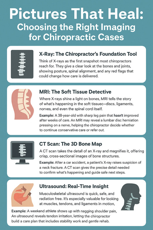
X-Ray: The Chiropractor’s Foundation Tool
Think of X-rays as the first snapshot most chiropractors reach for. They give a clear look at the bones and joints, showing posture, spinal alignment, and any red flags that could change how care is delivered.
When it’s helpful:
-
Spotting misalignments, scoliosis, or postural issues
-
Ruling out fractures, advanced osteoporosis, or tumors
-
Providing a baseline to track changes over time
Example: A patient comes in with long-standing low back pain and a history of falls. An X-ray can quickly show whether there’s arthritis, disc space narrowing, or something more serious that needs a cautious approach.
MRI: The Soft Tissue Detective
Where X-rays shine a light on bones, MRI tells the story of what’s happening in the soft tissues—discs, ligaments, nerves, and even the spinal cord itself.
When it’s helpful:
-
Finding herniated discs or nerve compression
-
Investigating unexplained numbness, weakness, or radiating pain
-
Checking on patients after surgery or when symptoms don’t improve
Example: A 38-year-old with sharp leg pain that hasn’t improved after weeks of care. An MRI may reveal a lumbar disc herniation pressing on a nerve, helping the chiropractor decide whether to continue conservative care or refer out.
CT Scan: The 3D Bone Map
A CT scan takes the detail of an X-ray and magnifies it, offering crisp, cross-sectional images of bone structures.
When it’s helpful:
-
Evaluating complex fractures or unusual bone shapes
-
Creating 3D views of the spine for clearer understanding
-
Checking whether bone is pressing into the spinal canal or nerve spaces
Example: After a car accident, a patient’s X-ray raises suspicion of a neck fracture. A CT scan gives the precise detail needed to confirm what’s happening and guide safe next steps.
Ultrasound: Real-Time Insight
Musculoskeletal ultrasound is quick, safe, and radiation-free. It’s especially valuable for looking at muscles, tendons, and ligaments in motion.
When it’s helpful:
-
Spotting rotator cuff injuries, sprains, or tendinopathy
-
Tracking how an injury is healing over time
-
Assessing extremity problems without needing advanced scans
Example: A weekend athlete shows up with nagging shoulder pain. An ultrasound reveals tendon irritation, letting the chiropractor build a care plan that includes stability work and gentle rehab.
Digital Motion X-Ray (DMX): Capturing Movement
Unlike regular X-rays, DMX shows the spine in motion, almost like a live video. It’s especially useful when pain comes from instability rather than something visible on a still image.
When it’s helpful:
-
Detecting ligament injuries and abnormal motion
-
Documenting whiplash or post-trauma instability
-
Supporting evidence in personal injury or workers’ comp cases
Example: A patient with lingering neck pain after a car accident has normal static X-rays. With DMX, subtle instability during flexion and extension becomes visible—explaining symptoms that otherwise seemed mysterious.
Why It Matters
Every image tells a story. For chiropractors, the key is knowing which story to ask for. Sometimes a simple X-ray is enough to confirm alignment and rule out danger. Other times, advanced imaging like MRI or CT uncovers issues that change the entire course of care.
By choosing the right tool at the right moment, chiropractors can:
-
Make safer, more accurate diagnoses
-
Build care plans rooted in clear evidence
-
Earn patients’ trust through transparency
-
Collaborate effectively with other healthcare providers when referral is needed
In the end, radiology isn’t about the machine—it’s about the message. The right picture doesn’t just show what’s wrong; it lights the way toward healing.